

Digital Preservation of the Vaquita Skeleton
Scientists have developed a digital reconstruction of the vaquita, the world’s most endangered marine mammal, to preserve its anatomy in three dimensions. This effort aims to support research and conservation initiatives as the species faces imminent extinction.
The project involved digitising the skeleton of a female vaquita, a species found exclusively in Mexico’s northern Gulf of California. Researchers employed a combination of medical imaging techniques, ultra-high-resolution micro CT scans, and photography to capture detailed anatomical data.
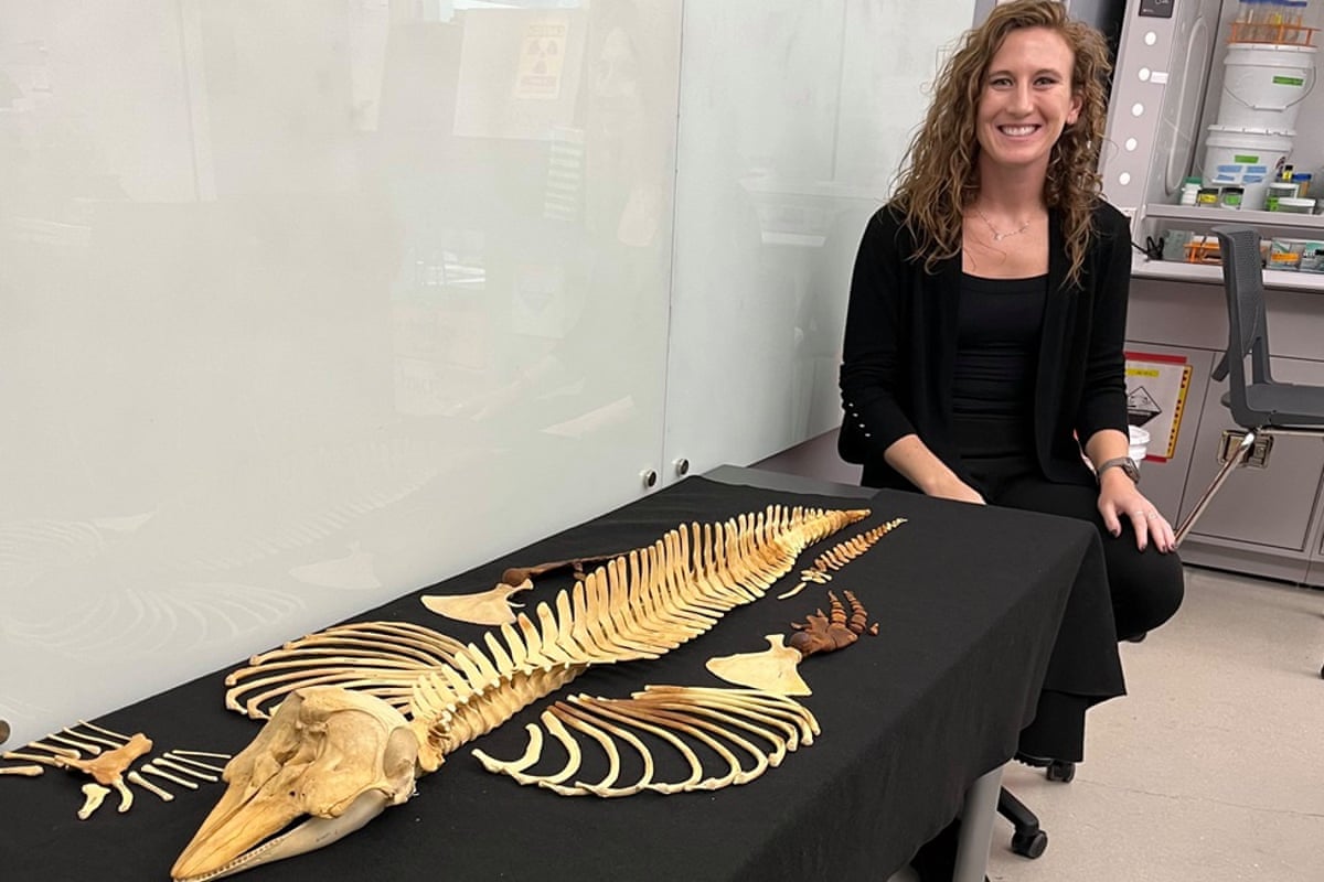
To facilitate global scientific study without risking damage to the rare and fragile physical specimens, the imaging data has been made freely accessible online. Only a few vaquita skeletons are believed to exist, making this digital archive particularly valuable.
“We want to influence conservation and awareness of the vaquita, but what it boils down to is open access datasets for biodiversity.
“There’s this whole web [of information] that can be shared to study biodiversity, conservation, evolution – there’s so many things that can come from one dataset.”
These remarks were made by Jamie Knaub, the study’s lead author and a doctoral researcher at Florida Atlantic University.
Population Decline and Conservation Status
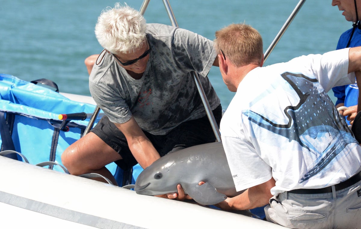
Historical estimates reported approximately 600 vaquitas in the wild. However, current figures from the World Wildlife Fund (WWF) suggest fewer than 10 individuals remain, establishing the vaquita as the rarest marine mammal on Earth.
The species’ decline is primarily attributed to bycatch in gillnets used by illegal fisheries targeting the totoaba, a large fish whose swim bladder is highly valued on international black markets.

Research Collaboration and Methodology
The research team, comprising members from Florida Atlantic University, the San Diego Natural History Museum, SeaWorld California, and NOAA Fisheries, based their work on a complete female vaquita skeleton collected in 1966.
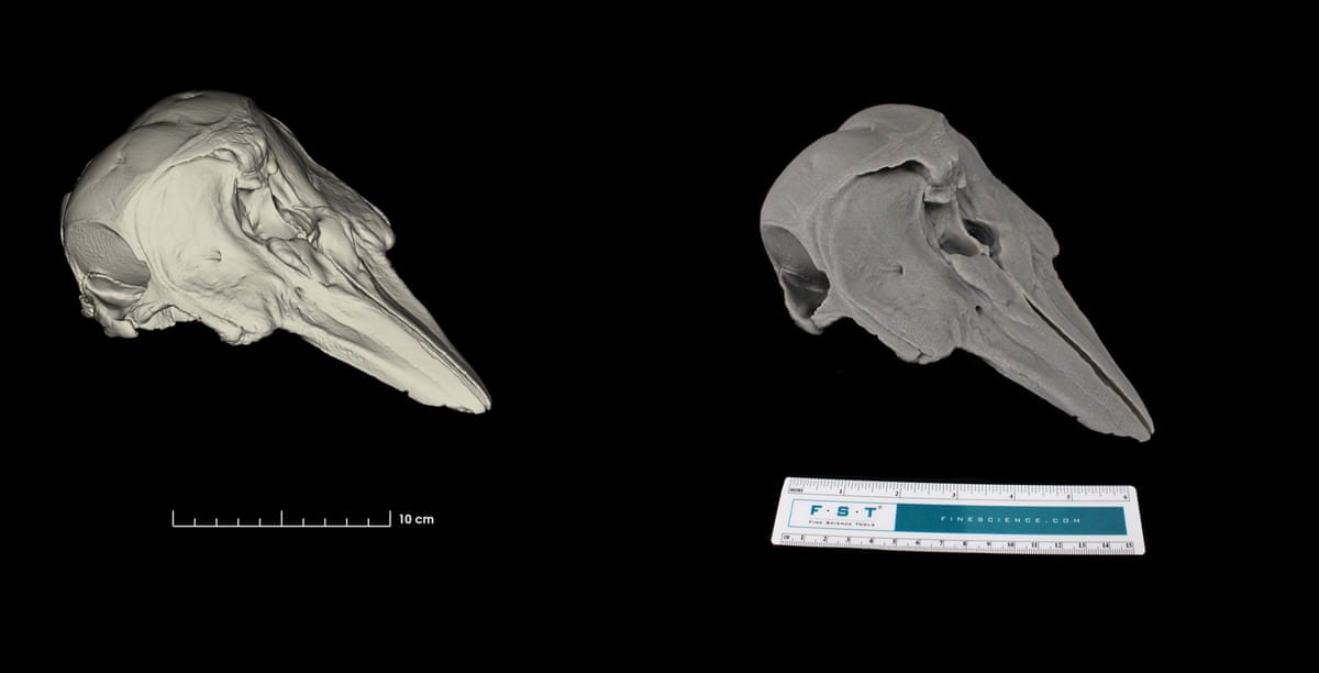
The study, published in a scientific journal, integrated hospital-grade CT scanning with microscopic CT imaging capable of resolving structures smaller than the width of a human hair. Thousands of scan slices were compiled to create three-dimensional models of every bone.
This methodology enabled the production of a highly detailed model, encompassing the entire skeleton down to microscopic bone structures.
Accessibility and Educational Applications
Due to the scarcity of vaquita skeletons, physical access is extremely limited. Knaub noted that the digital imaging could facilitate the creation of precise replicas for museum exhibits and educational settings, thereby increasing public awareness of the species.
Recent advances in imaging technology have accelerated efforts to digitise museum collections. Initiatives in the United States and elsewhere aim to make rare specimens accessible to researchers worldwide, eliminating the need to rely solely on photographs or to obtain permission to handle delicate originals.
“There’s a lot of people who don’t have access to museum specimens, or museums are wary of loaning out specimens because of how fragile or rare they are,”
Knaub said.
Species Description
The vaquita was only formally described in 1958. Growing to approximately 5 feet in length, it is the smallest member of the whale, dolphin, and porpoise family. It is distinguished by dark markings around its eyes and mouth.
For further information, and to follow biodiversity reporters, readers can access additional nature coverage through app.


